
Every practitioner who’s needled LI4 knows there isn’t just one sensation — there’s a quick surface prick, and then, deeper, something else: a heavy, spreading, dull ache.
Those two sensations aren’t the same event happening twice. They come from two distinct anatomical territories that the needle passes through on its way in, each with its own nerve supply, its own physiology, and — as it turns out — its own separate clinical relevance.

If you’re looking for more point actions, combinations and clinical uses for LI-4, click on LI-4 Acupuncture Point.
The Superficial Layer: Radial Nerve Territory and the Vascular Branch to the Radial Artery
The skin over the LI4 web space belongs entirely to the superficial branch of the radial nerve (SBRN) — sensory only. This is the initial prick when the needle breaks skin.
Umemoto et al. (2019), in Evidence-Based Complementary and Alternative Medicine, dissected 40 cadaver hands and traced vascular branches (VBs) of the SBRN, finding a VB reaching the radial artery in every specimen, closest to LI4 among the four points tested (LI2, LI3, LI4, LI5).
One caveat worth flagging: the study located LI4 using the traditional Japanese/Korean textbook convention (base of the metacarpals) rather than the WHO Standard location used throughout this article (midpoint of the 2nd metacarpal) — a discrepancy the authors clarified in a 2021 follow-up comment, attributing it to differing national conventions that predate WHO standardization.
The general finding — a vascular branch of the SBRN running through this region and connecting to the radial artery — likely still holds, but the specific distances reported (including the reported closeness relative to LI2/LI3/LI5) were measured from that alternate reference point, not the point this article describes.
This gives LI4’s general region a plausible anatomical basis for its classical use in peripheral circulatory conditions like Raynaud’s disease.
Independent support: Agarwal-Kozlowski, Lange, & Beck (2009), in a randomized, placebo-controlled crossover trial in Anesthesiology, used infrared thermography and found genuine deqi at LI4 significantly increased skin temperature, while sham needling decreased it — real physiological evidence for a circulatory effect at LI4 itself, independent of the exact anatomical distance question above.
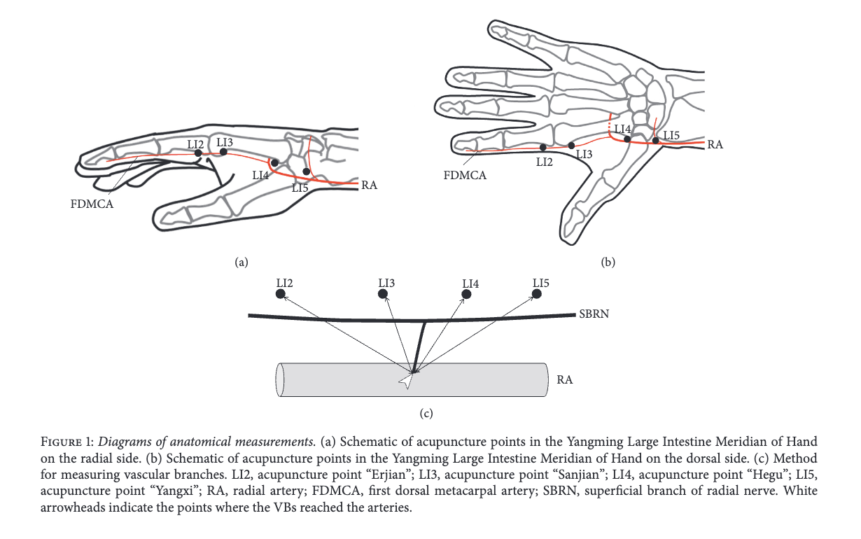
Figure adapted from Umemoto, K., Naito, M., Tano, K., et al. (2019). Acupuncture Point “Hegu” (LI4) Is Close to the Vascular Branch from the Superficial Branch of the Radial Nerve. Evidence-Based Complementary and Alternative Medicine, 2019, 6879076. Used under CC BY 4.0.
Note the proximal location of LI-4 for this study. This study was done by Korean scientists so the location is based more on Korean/Japanese LI-4 location – not TCM Acupuncture location.
The Deep Layer: Muscle Tissue and the Ulnar Nerve’s Motor Territory
Needle further and you reach the first dorsal interosseous (FDI) — the muscle belly that fills the space between the thumb and index finger metacarpals, and the structure LI4 sits directly within.
Here’s the anatomically well-established but counterintuitive part: despite sitting in “radial nerve territory” for skin sensation, the FDI’s motor supply comes entirely from the deep branch of the ulnar nerve.
The ulnar nerve enters the hand at the wrist through Guyon’s canal — on the pinky side — then its deep branch dives into the floor of the palm and travels the full width of the hand beneath the flexor tendons before terminating at the FDI. This is standard hand anatomy, not something specific to acupuncture research.
What this does and doesn’t explain. It’s tempting to conclude the ulnar nerve is what produces the deep ache — but no study actually shows this (that I could locate, anyway). Yang et al. (2013), reviewing the physiology of deqi in Evidence-Based Complementary and Alternative Medicine, describe a tissue-layer effect, not a nerve-specific one: stimulating muscle tissue — any muscle, regardless of which nerve innervates it — recruits the slower-conducting Aδ (delta) and C fibers responsible for aching, soreness, distension, and heaviness (the qualities that define deqi). Stimulating nerve branches directly tends to produce numbness via faster Aβ fibers instead. Stimulating blood vessels produces pain. This lines up with Kuo, Lin, & Ho (2004), who used laser Doppler flowmetry to characterize deqi specifically at LI4 and described it, in the subjects’ own words, as soreness and numbness — direct data from this exact point.
So the accurate statement is: the deep ache at LI4 comes from reaching muscle tissue — a depth and tissue-type effect that would occur at any sufficiently deep muscular point — and the muscle you happen to reach at LI4 is ulnar-innervated. The ulnar nerve is a real anatomical detail worth knowing, but it isn’t established as the mechanism behind the ache itself.
There’s also a local biochemical layer to this. Wu, Xu, Bai, Cui, Shu, He, et al. (2015), in the Journal of Chemical Neuroanatomy, examined cutaneous nerve terminal and mast cell responses specifically in the LI4 area of rats after manual acupuncture — adding a local biochemical dimension (neuropeptide and mast cell activity) beyond fiber conduction alone.
LI4’s Distinctive Signature in Neuroimaging
Separately from the fiber-type mechanism, LI4 shows up repeatedly in acupuncture neuroimaging research as producing a stronger-than-average response.
Kong et al. (2002), in the Journal of Alternative and Complementary Medicine, used fMRI to compare manual and electroacupuncture stimulation at LI4 and found differential brain activation between the two methods. Chen, Shen, Guan, Xiao, Du, & Wu (2011) ran an independent fMRI study specifically on manual-acupuncture-induced deqi at Hegu (LI4), finding activation in the prefrontal area, thalamus, striatum, and insular lobe — a second, independent line of neuroimaging evidence for LI4’s distinctive central response.
Broader review work (Yang et al., 2013) describes deqi at LI4 evoking deactivation of a limbic-paralimbic-neocortical network alongside activation of somatosensory regions — a distinct pattern from sharp pain, which activates rather than deactivates these same limbic areas. That same review also describes a PET imaging finding (Hsieh et al., as summarized within Yang et al.’s review) showing that eliciting deqi specifically at LI4 significantly increased blood flow to the hypothalamus and insula, extending into the midbrain — a secondhand citation, not one I’ve verified directly against Hsieh et al.’s original paper. LI4 shows a more prominent response than several other commonly studied points in these comparisons — a plausible partial explanation for why it’s reached for so often in practice, though the review doesn’t identify a single anatomical reason why.
The Electric Shock: A Different Sensation, Not a Stronger Deqi
Given how nerve-dense this small area is — sensory radial branches, autonomic vascular branches, and the ulnar nerve’s deep motor branch all in close proximity — it’s worth being precise about a sensation that’s easily confused with strong deqi: the sudden, sharp, instantaneously radiating “electric shock.”
Guo et al. (2020), in Neural Plasticity, make a well-supported case that this is a categorically different sensation from deqi, not an intensified version of it. Classical texts describe deqi as slow and gradually spreading; the electric shock is instantaneous and travels in a fixed direction down a limb.
Among major acupuncture sensation scales (see the Guo article for more info on these sensation scales), only two include the electric shock at all — three others exclude it specifically because of disagreement over whether it belongs in the category of normal needling sensation.
More concretely, Guo et al. reviewed documented case reports where the electric shock accompanied confirmed peripheral nerve injuries (median, peroneal, and sciatic nerve cases), each following continued or intensified needle manipulation after the shock occurred.
This distinction also has independent support from neuroimaging, not just case-report reasoning: Asghar, Green, Lythgoe, Lewith, & MacPherson (2010), in Brain Research, used fMRI to directly compare deqi and pain sensation and found genuinely different neural correlates for each.
Practical takeaway for LI4 specifically: a heavy, dull, spreading ache is consistent with genuine deqi in the muscle layer. A sharp, shooting, radiating sensation is a different category of event and — per both the case-report and neuroimaging evidence — reasonably treated as a signal to back the needle off slightly rather than a sign of deeper therapeutic engagement.
I often tell patients that if a shock is experienced (somewhat common in points like PC6 with the median nerve close by) — the sensation should disappear immediately and no further stimulation will be necessary. You could damage the nerve if you continued with stimulation but I can’t imagine a patient being able to tolerate this — let alone, a practitioner who is so willfully absent to a patient’s well-being.
The β-Endorphin Finding — and Why the Usual Framing Overstates It
A separate line of research examines LI4’s role in the classic acupuncture-analgesia pathway: opioid release.
Setiawardhani et al. (2017), in Journal of Physics: Conference Series (proceedings from a medicine and dentistry symposium), ran a small randomized trial: 18 subjects received 30 minutes of electroacupuncture at LI4, 18 received sham stimulation at a non-point, and plasma β-endorphin was measured before and after.
The result is commonly summarized as “EA at LI4 increases β-endorphin,” but the data tells a more specific story. The intervention group’s own before-after change was not statistically significant (34.6 → 35.1 pg/ml, p=0.897). What was significant is that the control group’s levels dropped sharply (43.6 → 10.3 pg/ml, p=0.001) while the LI4 group held steady. The accurate reading is that real stimulation at LI4 prevented a decline seen with sham treatment — not that it drove levels up. It’s still a real and interesting finding (and consistent with a genuine opioid-pathway effect), just worth stating precisely rather than with the more dramatic framing the abstract uses. Worth noting too: this is a conference proceedings venue rather than a dedicated clinical journal, with a small single-session sample — a real signal, not a settled finding.
There’s a systemic angle to this beyond the opioid pathway alone. Yu & Jones (2013), in a randomized, placebo-controlled trial published in Acupuncture in Medicine, examined electroacupuncture at LI4 (paired with LI11) and found that the intensity of deqi correlated directly with increases in sympathetic nervous system activity — measurable changes in heart rate, mean arterial pressure, and heart rate variability. This suggests LI4’s effects extend beyond the local site and beyond pain modulation alone, into a genuine autonomic/systemic response tied to how strongly deqi is elicited.
Clinical Synthesis
- For circulatory presentations (cold hands, Raynaud’s-type symptoms): the superficial vascular layer — SBRN’s proximity to the radial artery — is the relevant target. Strong deqi may not be necessary here; the mechanism implicated is vascular/autonomic, not muscular.
- For pain and deqi-dependent applications: deeper needling into the FDI muscle belly is what recruits the Aδ/C fiber response responsible for therapeutic deqi. The ulnar innervation of that muscle is a real anatomical fact, but not itself the mechanism — depth and tissue type are what matter.
- For systemic/autonomic effects: deqi intensity at LI4 appears to correlate with sympathetic nervous system activation — worth keeping in mind for presentations beyond local pain or circulation.
- Safety, at any depth: a sharp, radiating, electric-shock sensation is a distinct category from deqi — supported by both case-report and neuroimaging evidence — and a reasonable signal to ease off rather than intensify manipulation, especially relevant at LI4 given how much nerve tissue (sensory, autonomic, and motor) sits in this small area.
References
Agarwal-Kozlowski, K., Lange, A.-C., & Beck, H. (2009). Contact-free infrared thermography for assessing effects during acupuncture: a randomized, single-blinded, placebo-controlled crossover clinical trial. Anesthesiology, 111(3), 632–639. https://doi.org/10.1097/ALN.0b013e3181b31e24
Asghar, A. U. R., Green, G., Lythgoe, M. F., Lewith, G., & MacPherson, H. (2010). Acupuncture needling sensation: the neural correlates of deqi using fMRI. Brain Research, 1315, 111–118. https://doi.org/10.1016/j.brainres.2009.12.019
Chen, F., Shen, Z., Guan, J., Xiao, Y., Du, L., & Wu, R. (2011). Observation of the relation between brain activity and de qi sensation with manual acupuncture at LI4 (Hegu). Chinese Journal of Magnetic Resonance Imaging, 2(2), 112–117. DOI: 10.3969/j.issn.1674-8034.2011.02.008
Guo, Y., Zhu, K., Guo, J., Kuang, Y., Zhao, Z., & Li, W. (2020). The Electric Shock during Acupuncture: A Normal Needling Sensation or a Warning Sign. Neural Plasticity, 2020, 8834573. https://doi.org/10.1155/2020/8834573
Kong, J., Ma, L., Gollub, R. L., Wei, J., Yang, X., Li, D., Weng, X., Jia, F., Wang, C., Li, F., Li, R., & Zhuang, D. (2002). A pilot study of functional magnetic resonance imaging of the brain during manual and electroacupuncture stimulation of acupuncture point (LI-4 Hegu) in normal subjects reveals differential brain activation between methods. Journal of Alternative and Complementary Medicine, 8(4), 411–419. https://doi.org/10.1089/107555302760253603
Kuo, T.-C., Lin, C.-W., & Ho, F.-M. (2004). The soreness and numbness effect of acupuncture on skin blood flow. American Journal of Chinese Medicine, 32(1), 117–129. https://doi.org/10.1142/S0192415X04001825
Setiawardhani, A. L., Srilestari, A., & Simadibrata, C. (2017). Electroacupuncture effect at the LI4 Hegu point on the plasma β-endorphin level of healthy subjects. Journal of Physics: Conference Series, 884, 012027. https://doi.org/10.1088/1742-6596/884/1/012027
Umemoto, K., Naito, M., Tano, K., Terayama, H., Koike, T., Ohmichi, M., Ohmichi, Y., Sakabe, K., & Nakano, T. (2019). Acupuncture Point “Hegu” (LI4) Is Close to the Vascular Branch from the Superficial Branch of the Radial Nerve. Evidence-Based Complementary and Alternative Medicine, 2019, 6879076. https://doi.org/10.1155/2019/6879076
Wu, M.-L., Xu, D.-S., Bai, W.-Z., Cui, J.-J., Shu, H.-M., He, W., et al. (2015). Local cutaneous nerve terminal and mast cell responses to manual acupuncture in acupoint LI4 area of the rats. Journal of Chemical Neuroanatomy, 68, 14–21. https://doi.org/10.1016/j.jchemneu.2015.06.002
Yang, X.-Y., Shi, G.-X., Li, Q.-Q., Zhang, Z.-H., Xu, Q., & Liu, C.-Z. (2013). Characterization of Deqi Sensation and Acupuncture Effect. Evidence-Based Complementary and Alternative Medicine, 2013, 319734. https://doi.org/10.1155/2013/319734
Yu, D. T. W., & Jones, A. Y. M. (2013). Physiological changes associated with de qi during electroacupuncture to LI4 and LI11: a randomised, placebo-controlled trial. Acupuncture in Medicine, 31(2), 143–150. https://doi.org/10.1136/acupmed-2012-010280